Hereditary myopathies
Mitochondrial encephalomyopathy
Mitochondria are often called the powerhouses of eukaryotic cells. Pyruvate produced by glycolysis in the cytoplasm enters the mitochondria and via several inter-related complex processes, ATPs are generated via the tricarboxylic (TCA) cycle, followed by an efficient electron transport system at the inner membrane of the mitochondria. Lipids (another source of energy) are present in the cytosol in the form of fatty acids. Fatty acids are activated by acyl-CoA synthetase to produce acyl-CoA, which is transported across the inner mitochondrial membrane via the carnitine shuttle. The acyl-CoA is converted to acetyl-CoA through beta oxidation within the mitochondrial matrix. Subsequently, acetyl-CoA is used for energy production via the TCA cycle and the electron transport system.
Mitochondria possess their own small mitochondrial DNA (mtDNA), which encodes some of their own proteins, whereas other mitochondrial proteins are encoded by nuclear DNA. The mtDNA is small in size with a relatively simple structure. Human mitochondrial DNA is approximately 16 kb in size. A single mitochondrion usually contains two to ten copies of its DNA, which may not all be the same. Furthermore, heteroplasmy (heterogeneity of mitochondria within a single cell) is common. The mtDNA is inherited exclusively from the mother because only the ovum (in contrast to the sperm) contains mitochondria. Therefore, conditions associated with mtDNA abnormalities show maternal inheritance. (Table 2)
Major mitochondrial encephalomyopathies
- Chronic progressive external ophthalmoplegia ( CPEO )
- Kearns-Sayre syndrome ( Onset before age 20 ,CPEO, retinitis pigmentosa, cardiac conduction block, cerebellar ataxia )
- Frequently associated with deletion in mtDNA
- Mitochondrial myopathy, encephalopathy, lactic acidosis and stroke-like episodes ( MELAS )
- Associated with point mutation of mtDNA3,243, or 3,271. Maternal inheritance.
- Myoclonus epilepsy associated with ragged-red fiber ( MERRF )
- Associated with point mutation at mtDNA8,344, Maternal inheritance.
- Leigh encephalopathy
- Subacute necrotizing encephalomyelitis in the basal ganglia, brain stem, thalamus and posterior column of the spinal cord.
- Muscle weakness, delayed mental and motor development in infants.
- Maternal, AR of XR inheritance. Many mutations in both mtDNA and nuclear DNA have been reported.
Tab.2
1. Kearns-Sayre syndrome and chronic progressive external ophthalmoplegia
Chronic progressive external ophthalmoplegia (CPEO) refers to chronic progressive difficulty in eye movements. Kearns-Sayre syndrome (KSS) is a mitochondrial disease characterized by CPEO and other features such as hearing difficulty, retinitis pigmentosa, mental retardation, cerebellar ataxia, cardiac conduction defects, and diabetes mellitus. Pearson syndrome refers to early-onset sideroblastic anemia and exocrine pancreatic dysfunction.
More than 50% of patients with CPEO and most patients with KSS and Pearson syndrome show a large deletion in mtDNA. Most patients with a large deletion in mtDNA show a single mutation; however, multiple heterogeneous mutations may occur in a few cases. The latter condition is interpreted as impairment of DNA repair or reproduction and may be attributed to mutations in the nuclear DNA showing Mendelian inheritance. A few reports have described CPEO caused by mutations in nuclear DNA. AD progressive external ophthalmoplegia is known to occur secondary to mutations in genes such as the polymerase gamma (POLG1 and 2) and several other nuclear genes.
Myopathological findings include ragged red fibers (RRF) and fibers with low cytochrome C oxidase (COX) activity. RRFs show a wide range of COX activity varying from low to high (Fig. 31).
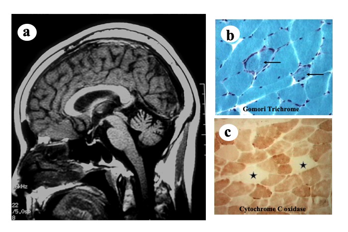
Fig.31
Kearns-Sayre syndrome: A patient with CPEO, cerebellar ataxia with muscle weakness. Head MRI shows cerebellar atrophy (a), and muscle biopsy shows ragged red fibers (b: arrows), and fibers with low activity of cytochrome C oxidase (c: stars).
Electron microscopy shows mitochondrial paracrystalline inclusions (Fig. 32).
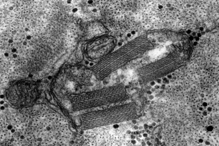
Fig.32
Electron micrograph of paracrystlline inclusion of mitochondria.


