Hereditary myopathies
Muscular dystrophies
2. Becker muscular dystrophy (X-linked recessive muscular dystrophy, Becker type)
(1) Pathophysiology
Becker muscular dystrophy (BMD) is characterized by weaker-than-normal expression of dystrophin, although it is not completely absent as noted in patients with DMD. This is because despite the mutation, the reading frame is preserved and allows the production of dystrophin that is abnormal in quantity and function. Western blot analysis of dystrophin in patients with BMD reveals only 35–80% of the normal quantity of dystrophin, and it is of a lower-than-normal molecular weight.
(2) Symptoms and signs
Clinical features of BMD vary greatly. BMD and DMD often show a similar clinical presentation in a few patients, whereas a few patients are nearly asymptomatic. Severe cases are often referred to as “outlier” of DMD. Asymptomatic cases can be misdiagnosed as idiopathic hyper-CKemia. Usually, clinical features mimic those observed in patients with limb-girdle muscular dystrophy (LGMD). Symptom onset occurs before 10 years of age. Relatively slow progression of the disease results in inability to walk by the fourth decade of life. Symptoms resemble those of DMD but are milder in severity. Pseudohypertrophy of the calves is commonly noted. Cardiomyopathy and intellectual retardation are uncommon but may occur.
(3) Histopathological and immunohistochemical examinations
Basic histopathological changes resemble those observed in cases of DMD, although these are less severe. Immunohistochemical analysis of dystrophin shows various patterns depending on the epitope of the antibody and location of the mutation in the DMD gene. The usual pattern consists of weakly stained dystrophin along with various degrees of unevenness (Fig. 17).
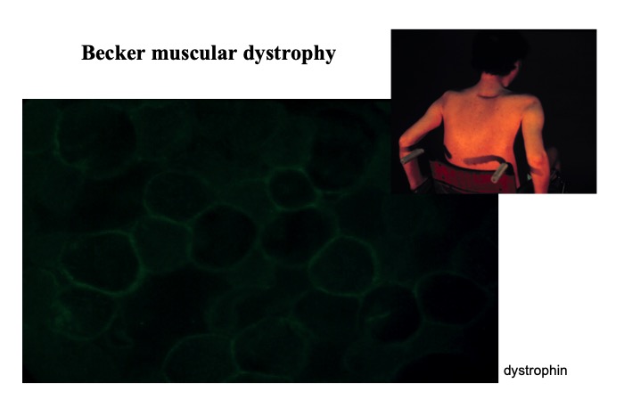
Fig.17
Dystrophin in Becker muscular dystrophy showing weak and uneven positivity.
However, totally unstained fibers are relatively few. Patients with mild clinical symptoms are often difficult to diagnose based on genetic and histopathological evaluation. In such cases, two or three different antibodies against different epitopes need to be used along with antibodies against other membrane proteins. Genetic analysis and quantitative estimation of dystrophin with a Western blot study is also useful.


