Hereditary myopathies
Congenital myopathies
3. Centronuclear myopathy
In more than 95% of mature normal skeletal muscle fibers, the nuclei are present near the surface of the fiber. However, the occurrence of a central or internal nucleus (myonuclei at the center of the sarcoplasm) is significantly high in patients with myopathies and chronic neurogenic atrophy. Therefore, the presence of an internal nucleus is a non-specific change. Centronuclear myopathy is a congenital myopathy characterized by significantly increased numbers of internal nuclei, and this histopathological feature predominates over all other changes (Fig. 28). Rouleaux formation-like long chain of stacked internal nuclei are commonly noted in longitudinal sections of muscle fibers.
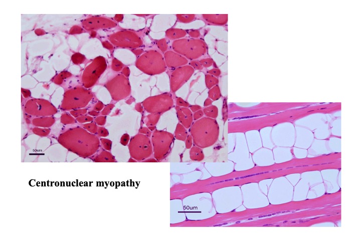
Fig.28
Centronuclear myopathy is characterized by high frequency of internal nuclei. The central nuclei are often observed in rows mimicking Rouleaux formation-like chains.
Centronuclear myopathy is caused by mutations in at least the following six genes: myotubularin (MTM 1) gene mutations cause the most severe infantile X-linked myotubular myopathy, dynamin 2 (DNM2) gene mutations are associated with an AD early-onset centronuclear myopathy, amphiphysin 2 (BIN1) gene mutations are associated with an AD or AR juvenile onset disease, the coiled-coil domaincontaining protein 78 (CCDC78) gene mutation is associated with an AR juvenile form of disease, and mutations in the RYR1 and TTN genes can cause AR centronuclear myopathies.
As described earlier, centronuclear myopathies are closely related to many other congenital myopathies. Although the exact mechanism of formation of the central nucleus is unclear, several genes associated with central nuclei are known to code proteins that occur in the sarcoplasmic reticulum, transverse systems, or the nuclei and are closely involved in transport of materials, as well as in autophagy (Jungbluth, 2014).


