Muscle biopsy
Normal muscle
A fascicle is a bundle of muscle fibers, which appear polygonal in shape on a transverse section. The fibers show a cobblestone (paving stone) appearance with little space between them (Fig. 1, 2).
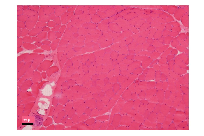
Fig.1
Normal muscle (frozen section, HE stain)
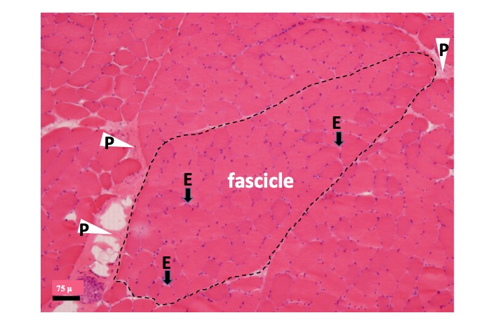
Fig.2
A fascicle is surrounded by interstitial tissue called perimysium (P) and consists of tightly packed muscle fibers with narrow, barely visible interstitial tissue called endomysium (E).
Most muscle fibers show peripheral nuclei. A nucleus present at the middle of the sarcoplasm (cytoplasm of the muscle cell) on transverse section is called a central or internal nucleus. Normal skeletal muscle shows internal nuclei in ≤3% of all fibers. Significant age-related variations are noted in muscle fiber size. In healthy adults, most fibers measure 40–60 μm in diameter.
The narrow interstitial space between muscle fibers within the fasciculus is called endomysium, and the wider interstitial tissue between the fasciculi is called perimysium. The perimysium contains blood vessels, fat, intramuscular nerve fasciculi, and muscle spindles (Fig. 3).
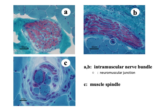
Fig.3
In the perimysium, blood vessels, fatty tissue, intramuscular nerve bundles (a, b) and muscle spindles (c) are observed. In the vicinity of the intramuscular nerved fibers, neuromuscular junctions can be seen occasionally (c).
Determination of the fiber type is an important component of histochemical analysis of muscle tissue (Fig. 4).
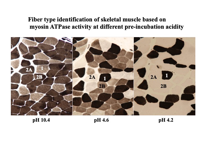
Fig.4
Healthy mature skeletal muscle contains a mixture of type 1 and 2 fibers. Type 2 fibers are further categorized as type 2A and type 2B. Type 1 fibers are referred to as slow red muscle fibers, which depend on aerobic glycolysis for energy production and are resistant to fatigue on continuous contraction. Type 2 fibers are referred to as fast white muscle fibers. Type 2A fibers rely on aerobic and anaerobic glycolysis for energy and are relatively more resistant to continuous exercise than type 2B fibers, which rely exclusively on anaerobic glycolysis for energy and show easy fatigability on continuous exercise.
Electron microscopy reveals the ultrastructure of skeletal muscle (Fig. 5).
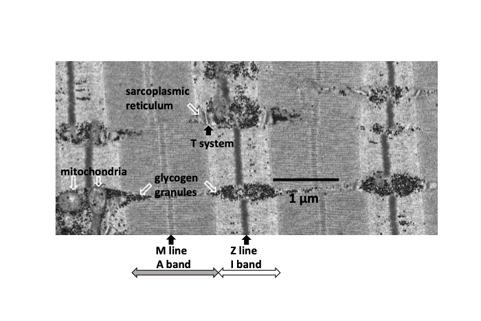
Fig.5
Electron micrograph of normal skeletal muscle
A muscle fiber contains myofibrils, which contain two types of myofilaments, the thin and thick myofilaments. The thin myofilaments are mainly composed of actin, whereas thick filaments are mainly composed of myosin. Z-disks and M lines hold the myofilaments in place and maintain the structural arrangement and layering and order of myofibrils. The thin filaments are fixed at the Z-disks. Contraction of the muscle fiber involves a specific sequence of events that begin with the signal transmitted from the motor neuron innervating that fiber (the role of acetylcholine has been explained earlier). Following the action of calcium and sodium ions at the level of the muscle fiber membrane and other processes, the thin filaments slide into the thick filaments, which are pulled close to each other at the M lines and this activity initiates contraction of the muscle. The sarcomere is the actual functional (contractile) unit of the muscle fiber and represents the region containing myofilaments between two Z-disks. In addition to myofibrils, it contains the transverse systems (T-systems), sarcoplasmic reticulum, and mitochondria. At the surface of the muscle fiber is the plasma membrane, which is covered by a basal lamina. Muscle cell nuclei (myonuclei) are observed beneath the surface of fibers and are accompanied by the Golgi apparatus and glycogen granules. Satellite cells and neuromuscular junctions can be visualized on the surface of muscle fibers (Fig. 6).
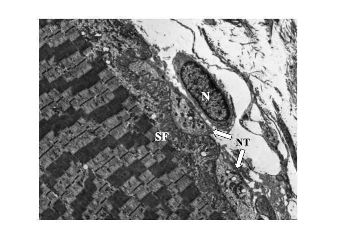
Fig.6
Ultrastructure of neuromuscular junction
N: nucleus of a Schwann cell, NT: nerve terminal, SF: synaptic folds


