⑫ Staining
Klüver-Barrera(KB)staining
Double staining with Nissl staining using cresyl violet and LFB staining of myelin sheaths.
See the Nissl and LFB staining sections for more detailed information on these methods.
Staining procedure
| 1 | 1 Deparaffinization | Xylene | 3 changes, 10 minuntes each |
| 2 | Removal of xylene | 100% ethanol | 3 changes, 5 minutes each |
| 3 | Hydration | 95% ethanol | 5 minutes |
| 4 | Staining | 0.1% luxol fast blue solution | Overnight, 50℃ |
| 5 | Rinsing | 95% ethanol | 1 minute |
| 6 | Washing | Running tap water | |
| 7 | Differentiation | 0.05% lithium carbonate solution | 5 seconds to 20 seconds |
| 8 | Differentiation | 70% ethanol | 3 changes. 1 minute each in the first and second dish. Leave sections in the third dish for approximately 1 to 10 minutes. |
| 9 | Microscopic check | | Repeat step ⑦&⑧, until gray matter become white. |
| 10 | Washing | Running tap water | |
| 11 | Rinsing | Distilled water | |
| 12 | Staining | 0.1% cresyl violet solution | 10 minutes, 37℃ |
| 13 | Differentiation | 95% ethanol + a few drops of 10% acetic acid solution | Approximately 5 to 10 minutes. Care should be taken not to overdifferentiate since decolorization will proceed in the following step |
| 14 | Differentiation | 95%, 100% ethanol | Approximately 1 minute each. Differentiate until only nuclei and nissl bodies are blue purple. |
| 15 | Microscopic check | | Step ⑦ & ⑧ may be repeated if more differentiation is needed. |
| 16 | Dehydration | 100% ehtanol | 2 changes, 5 minutes each |
| 17 | Clearing | Xylene | 3 changes, 10 minuntes each |
| 18 | Coverslipping | | |
Staining results
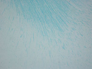
The color tones of neurons and myelin sheaths are more distinct with KB staining compared with LFB or Nissl staining alone. The change in color tone is particularly notable for the myelin sheaths. With LFB staining, the myelin sheath is stained emerald green, whereas it is stained navy blue with KB staining, which creates a sharper contrast.
Staining pattern
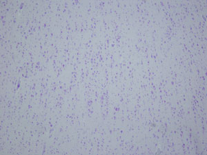
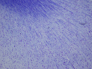
In a Loupe image, the gray matter is stained a pale, cloudy blue color and the white matter is stained bright blue, which improves the detection of white matter lesions.
Nerve cell body
| Normal histology |
Pathology |
| Nissl substance- blue purple | Achromasia - pale blue color with loss of nissle substance |
| Lipofuscin - yellowish brown |
| Neuromelanin - Dark brown |
Nerve process
| Normal histology |
Pathology |
| Unstained | Unstained |
Astrocyte
| Normal histology |
Pathology |
| Unstained | Rosenthal fiber-blue |
Oligodendrocyte
| Normal histology |
Pathology |
| Unstained | Unstained |
Myelin sheath
| Normal histology |
Pathology |
| The sheath wrapped around the axon-blue | Demyelinated areas are unstained. |
Macrophage
| Normal histology |
Pathology |
| Unstained | Myelin fragments recently engulfed by macrophages - blue |
| Fat granule cell - colorless |
| Iron granule cell - brown |