⑫ Staining
Nissl staining
Nuclei and nissl bodies (rough endoplasmic reticulum) are stained blue purple with cresyl violet.
Nissl staining is used for Klüver-Barrera (KB) staining.
Staining procedure
| 1 | Deparaffinization | Xylene | 3 changes, 10 minuntes each |
|---|---|---|---|
| 2 | Removal of xylene | 100% ethanol | 3 changes, 5 minutes each |
| 3 | Hydration | 95%, 70% ethanol | 5 minutes each |
| 4 | Washing | Running tap water | 2 minutes |
| 5 | Rinsing | Distilled water | |
| 6 | Staining | 0.1% cresyl violet solution | 10 minutes, 37℃ |
| 7 | Differentiation | 95% ethanol + a few drops of 10% acetic acid solution | Approximately 5 to 10 minutes. Care should be taken not to overdifferentiate since decolorization will proceed in the following step. |
| 8 | Differentiation | 95%, 100% ethanol | Approximately 1 minute each. Differentiate until only nuclei and nissl bodies are blue purple. |
| 9 | Microscopic check | Step ⑦ & ⑧ may be repeated if more differentiation is needed. | |
| 10 | Dehydration | 100% ehtanol | 2 changes, 5 minutes each |
| 11 | Clearing | Xylene | 3 changes, 10 minuntes each |
| 12 | Coverslipping |
0.1% cresyl violet aqueous solution (Filter before use)
| cresyl violet | 1g |
|---|---|
| distilled water | 1000ml |
pH of cresyl violet
The staining method differs depending on the pH of the staining solution. At a pH close to 3.0, only the Nissl bodies, nucleoli, and nuclear membranes are stained pale blue. Glial cell nuclei are only very slightly stained. As the pH increases, the color becomes darker overall, and the cytoplasm of nerve cells, nerve fibers, and glial cells are co-stained. Co-staining intensifies close to a pH of 4.0. Therefore, a longer differentiation time is required.
pH3.0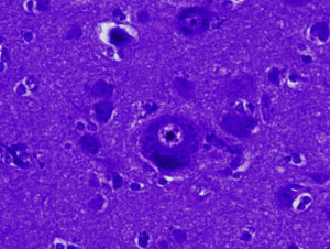
pH4.7
Types of cresyl violet
Three types of cresyl violet are currently used in the Laboratory of Neuropathology.
| product name | manufacturer | availability |
|---|---|---|
| Cresyl Violet Acetate | EM SCIENCE | out of stock |
| Cresyl Violet acetate | SIGMA | Available commercially |
| Cresyl violet (Acetate) | MERCK | Available commercially |
Differentiation time differs depending on the cresyl violet product
The pH of the staining solution differs depending on the product. Therefore, the staining methods also differ. It is important to be aware that the time required for differentiation differs between products. However, whichever product is used, the same stained chromatic image can ultimately be obtained through differentiation and observation of the specimen under a microscope.
| product name | manufacturer | pH of 0.1% cresyl violet solution | Differentiation time in 95% ethanol |
|---|---|---|---|
| Cresyl Violet Acetate | EM SCIENCE | 3.5 | for 10 to 30 seconds |
| Cresyl Violet acetate | SIGMA | 4.7 | 10min approximately |
| Cresyl violet (Acetate) | MERCK | 4.8 | 10min approximately |
※ The pH can be adjusted from 3.5 to approximately 3.8 by adding a suitable amount of 10% acetic acid to the Sigma and Merck staining solutions, which will shorten the time needed for differentiation with 95% ethanol. However, it is not recommended to reuse this solution.
Important considerations regarding differentiation
Perform differentiation until the background turns white. Having the nucleoli and nuclear membranes of the nerve cell nuclei clear and chromatin faintly stained is ideal.
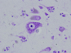
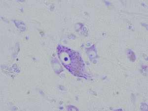
Cresyl Violet acetate (Sigma C5042) pH4.7 was used in the both sections.
Left photograph: 95% ethanol differentiation for only 10 seconds. With insufficient differentiation, the entire nerve cell is stained blue, making it impossible to identify the nucleus and Nissl bodies.
Right photograph: 95% ethanol differentiation for 10 minutes. Sufficient differentiation allows the nuclei and Nissl bodies of neurons to be clearly differentiated.