⑫ Staining
Luxol fast blue (LFB) staining
Myelin sheaths are stained blue with Luxol fast blue.
Luxol fast blue is used for Klüver-Barrera (KB) staining
Staining procedure
| 1 | Deparaffinization | Xylene | 3 changes, 10 minuntes each |
|---|---|---|---|
| 2 | Removal of xylene | 100% ethanol | 3 changes, 5 minutes each |
| 3 | Hydration | 95% ethanol | 5 minutes |
| 4 | Staining *1 | 0.1% luxol fast blue solution | Overnight, 50℃ |
| 5 | Rinsing | 95% ethanol | 1 minute |
| 6 | Washing *2 | Running tap water | |
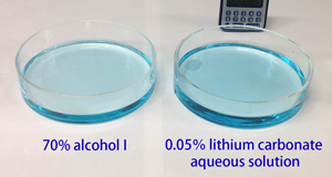
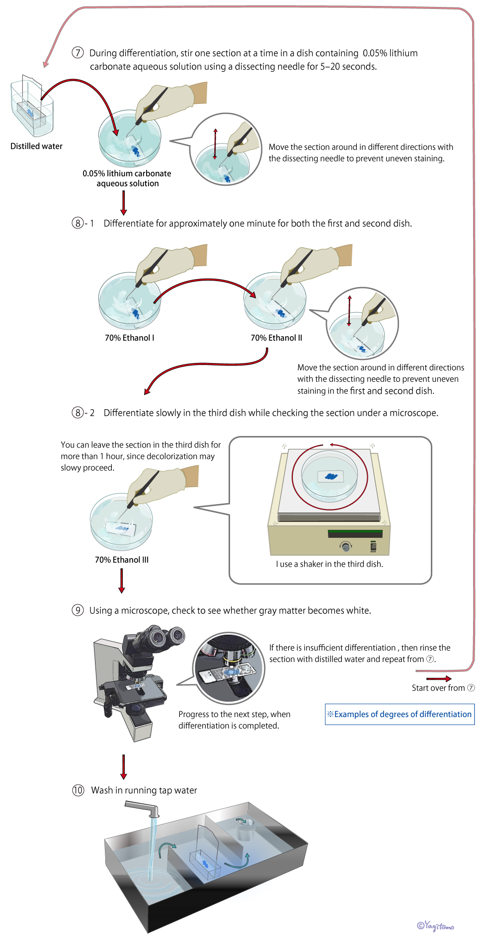
| 7 | Differentiation *3 | 0.05% lithium carbonate | 5 seconds to 20 seconds |
| 8 | Differentiation | 70% ethanol | 3 changes. 1 minute each in the first and second dish. Leave sections in the third dish for approximately 1 to 10 minutes. |
| 9 | Microscopic check | Repeat step ⑦ & ⑧, until gray matter become white. | |
| 10 | Washing | Running tap water | |
| 11 | Dehydration | 70%, 95% ethanol | 5 minutes each |
| 12 | Dehydration | 100% ehtanol | 3 changes, 5 minutes each |
| 13 | Clearing | Xylene | 3 changes, 10 minuntes each |
| 14 | Coverslipping |
0.1% Luxol fast blue solution (Filter before use)
| Luxol fast blue | 1g |
|---|---|
| 95% ethanol | 1000ml |
| 10% acetic acid solution | 5ml |
Types of LFB
Two types of LFB are currently used in the Laboratory of Neuropathology. Whichever product is used, the same stained chromatic image can ultimately be obtained through differentiation and observation of the specimen under a microscope.
| Product name | Manufacturer | Availability |
|---|---|---|
| Luxol fast blue MBSN (Solvent blue 38) | EM SCIENCE | Out of stock |
| Solvvent blue 38 (Luxol fast blue MBSN) | SIGMA | Available commercially |
Important considerations regarding LFB staining
*1 Staining
- After LFB staining, return the specimen to room temperature and progress to the next procedure.
*2 Washing
Differentiate one section at a time in a dish of the differentiating solution.
- Differentiate one section at a time in a dish of the differentiating solution.
- Stir the section in the solution with a dissecting needle.
*3 Differentiation
- Differentiate one section at a time in a dish of the differentiating solution.
- Stir the section in the solution with a dissecting needle.
- The first dish quickly becomes dirty. Therefore, replace it with a new solution when its color darkens.
- Leave the section in the third dish until gray matter become colorless.
- Shake the third dish during the process.
- Step ⑦ & ⑧ may be repeated if more differentiaion is needed.
- Too much or too little differentiation produces false positives and false negatives, respectively. Care should be taken during the process.
⑥ 〜 ⑨ Experiment video
⑥ 〜 ⑨ Commentary illustrations
Important considerations regarding differentiation
- If too much color is lost in step ⑥, then this is irreparable and differentiation must be carefully conducted. Slowly and thoroughly remove the color in step ⑦.
- Repeat steps ⑥–⑧ until the cortex turns white. During this process, take care to avoid removing more color than necessary, which would end up removing the main pathological structures.
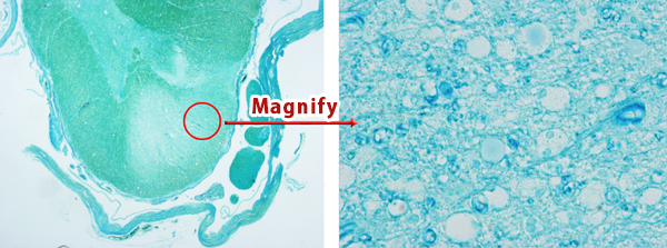
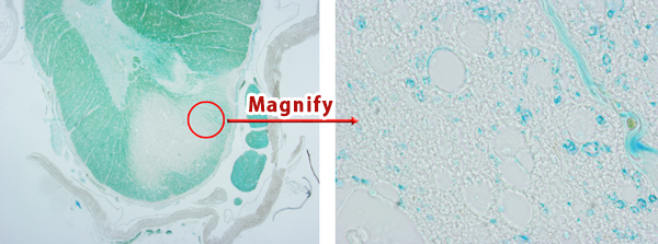
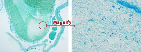
Examples of degrees of differentiation
Amyotrophic lateral sclerosis of the lateral funiculus of the spinal cord: The breakdown of the myelin sheath.
Overdifferentiation
The background is completely colorless, and it is difficult to find phagocytosis by macrophages.
Moderate differentiation
We can see the breakdown products of myelin sheathes engulfed by macrophages.
Underdifferentiation
The entire specimen is blue. It is impossible to distinguish the breakdown products of the myelyn sheath.