| Japanese | English |
|


Tissue diagnosis heavily depends on the quality of sections. Therefore, histotechnologists need skills to prepare sections that accurately reflect cell components and lesions.
We are committed to spread knowledge and skills of the precise preparation of sections in neurohistology.
An incorrect stain may lead to serious errors in diagnosis.
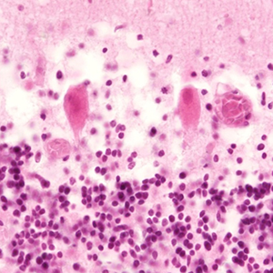
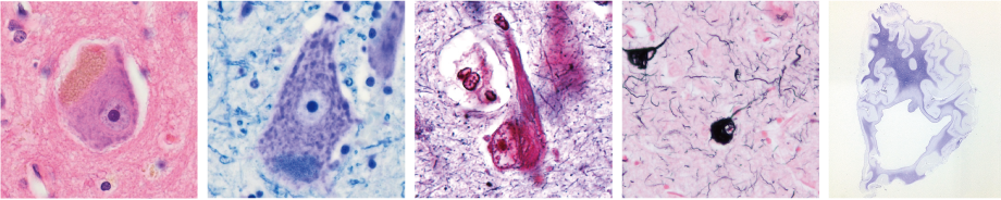
The figure on the left shows a hematoxylin and eosin-stained cerebellum specimen. Two Purkinje cells are shown in the center. When the cerebellum is stained with hematoxylin and eosin, normal Purkinje cell nuclei are stained blue purple and the cytoplasms are stained pink. However, the cytoplasms and nuclei of these two Purkinje cells appear deep red and atrophied.
Therefore, these Purkinje cells are not normal. Noting the reddened and atrophied cytoplasms, many people would diagnose these features as ischemic changes.
However, these are not ischemic changes. In fact, a degraded staining solution was used. Therefore, this result is due to the cells not achieving the correct color stain. This is an example of inappropriate staining leading to misdiagnosis.
In other words, appropriately performing staining is an important element of pathological diagnosis.

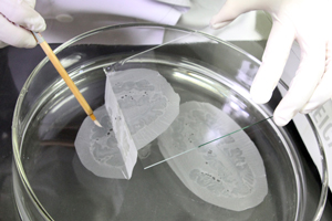
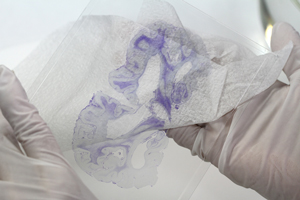
Preparing large brain sections
We are one of the rare laboratories preparing large brain sections in Japan.
The large brain section allows us to detect the distribution patterns of lesions in brain diseases.
It can be also compared with imaging tests (e.g., CT, MRI).
Staining methods for the central nervous system
Central nervous system has specialized structures compared to other tissues. These include normal components such as myelin sheaths and nerve processes. They can be detected using special staining methods.
We use a general staining method such as Hematoxylin and Eosin method to give an overview of tissue structures, and several neurohistological staining methods.

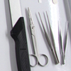
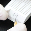
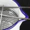
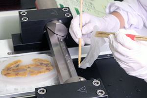
⑩ Sectioning
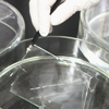
Procedure to thinly slice the tissue and affix it to a glass slide for staining and microscopy
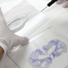
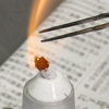
⑫ Staining
Procedure to thinly slice the tissue and stain its components with pigment to produce a specimen for optical microscopy
- ■ Hematoxylin and Eosin(HE)staining
- ■ Nissl staining
- ■ Luxol fast blue(LFB)staining
- ■ Klüver-Barrera(KB)staining
- ■ Woelcke staining
- ■ Bodian staining
- ■ Holmes staining
- ■ Holmes staining(Eguchi-Seki modification)
- ■ Gallyas-Braak(GB)staining
- ■ Holzer staining
- ■ Marchi staining
- ■ Berlin blue staining
- ■ Kossa staining
- ■ Azan staining
- ■ Immunohistochemical(IHC)staining
・ Enzymatic method
・ Immunofluorescence(IF)method - ■ Nuclear staining