⑫ Staining
Hematoxylin and Eosin(HE)staining
Hematoxylin stains the cell nucleus blue purple, whereas eosin stains other structures in various shades of red.
Staining procedure using Mayer’s hematoxylin solution
| 1 | Deparaffinization | Xylene | 3 changes, 10 minuntes each |
|---|---|---|---|
| 2 | Removal of xylene | 100% ethanol | 3 changes, 5 minutes each |
| 3 | Hydration | 95%, 70% ethanol | 5 minutes each |
| 4 | Washing | Running tap water | 2 minutes |
| 5 | Rinsing | Distilled water | |
| 6 | Staining | Hematoxylin solution | 4 minutes |
| 7 | Blueing | Running tap water | 15 minutes |
| 8 | Staining | Eosin solution | 2 minutes |
| 9 | Differentiation | 70% ethanol | 10 quick dips |
| 10 | Dehydration | 95% ethanol | Quickly |
| 11 | Dehydration | 100% ehtanol | 3 changes, 5 minutes each |
| 12 | Clearing | Xylene | 3 changes, 10 minuntes each |
| 13 | Coverslipping |
⑥ 〜 ⑫ Experiment video
Procedure
Staining solution
Mayer’s hematoxylin solution
| hematoxylin | 1.0g |
|---|---|
| distilled water | 1000ml |
| Sodium iodade | 0.2g |
| Either potassium alum or Ammonium alum | 50g |
| Chloral hydrate | 50g |
| Citric acid | 1.0g |
There are many types of hematoxylin solutions. The typical hematoxylin solutions are those of Mayer and Carracci.
Mayer’s staining solution contains acid, whereas Carracci’s hematoxylin is relatively neutral because it does not contain acid. Therefore, the staining methods differ between the Mayer and Carracci stains.
With Mayer’s hematoxylin, the nucleus is stained red purple at first and then turns blue purple under running water. Conversely, with Carracci’s hematoxylin, the entire tissue is stained blue purple and then differentiated with hydrochloric acid-alcohol solution, resulting in only the nucleus retaining the blue-purple stain.
Eosin solution
*1.0% eosin solution(stock solution)
| Eosin Y, water-soluble | 1.0g |
|---|---|
| distilled water | 100ml |
* Working solution of eosin
| 1.0% eosin solution | 20ml |
|---|---|
| 80% ethanol | 160ml |
| acetic acid | 10滴 |
Maintaining the quality of the staining solution
Hematoxylin
After preparing the staining solution, the staining results change as the stain is used multiple times. The following three explanations may be related to this change:
- The natural oxidation of hematoxylin.
- The staining solution becomes contaminated with water which is carried with a section.
- The concentration of the staining solution decreases as the pigment is consumed.
It is essential to regularly change the staining solution to maintain consistent staining results. Refrigerating the staining solution makes the solution last longer; however, the solution must be at room temperature when used.
Staining deterioration due to the natural oxidation of hematoxylin
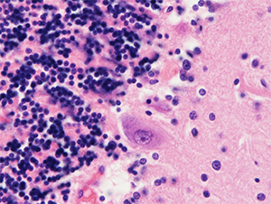 Fresh hematoxylin solusion
A cerebellum specimen stained immediately after preparing the hematoxylin. The color of the Purkinje and granule cell nuclei is blue purple.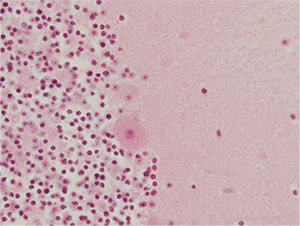 Old hematloxylin solusion
A cerebellum specimen stained one year after preparing the hematoxylin. The color of the Purkinje and granule cell nuclei is red purple.
Eosin solution
Unlike hematoxylin, eosin still stains well even when the solution is old.
When a stronger stain is needed, adding a small amount of acetic acid is effective.
Staining pattern
Please note that, although almost all normal and abnormal structures can be observed with HE staining, some structures that are important for differential diagnosis cannot be visualized using this staining method (see table below).
Therefore, these structures require special staining or immunostaining.
Basophilic appearance differs slightly depending on the hematoxylin solution used (e.g., Carracci or Mayer).
Nerve cell body
| Normal histology | Pathology | Structures invisible in H&E preparations | ||
|---|---|---|---|---|
| Eosinophilic |
|
Eosinophilic |
|
detected by TDP-43 and ubiquitin immunostaining
detected by polyglutamine immunostaining) |
| Basophilic |
|
Basophilic |
|
|
| Others |
|
|||
|
||||
Nerve process
| Normal histology | Pathology |
|
||
|---|---|---|---|---|
| Eosinophilic |
| Eosinophilic |
|
detected by gallyas-braak staining and tau immunostaining |
Astrocyte
| Normal histology | Pathology | Structures invisible in H&E preparations | ||
|---|---|---|---|---|
| Eosinophilic |
| Eosinophilic |
|
detected by tau - immunostaining and gallyas - braak staining |
| Basophilic |
| Basophilic |
|
|
Oligodendrocyte
| Normal histology | Pathology | Structures invisible in H&E preparations | ||
|---|---|---|---|---|
| Eosinophilic |
|
Eosinophilic |
|
|
| Basophilic |
| Basophilic |
|
|
Myelin sheath
| Normal histology | Pathology | ||
|---|---|---|---|
| Eosinophilic |
| Eosinophilic |
|
Microglia
| Normal histology | Pathology | ||
|---|---|---|---|
| Basophilic |
|
Basophilic |
|
Macrophage
| Normal histology | Pathology | ||
|---|---|---|---|
| Basophilic |
| Eosinophilic |
|
| Others |
| Others |
|
|
|||