In 1817, James Parkinson provided the first detailed description of the clinical pattern of Parkinson’s disease. In 1917, Tretiakoff’s thesis presented the clinicopathological aspects of the disease, showing that the responsible lesions were in the midbrain substantia nigra.
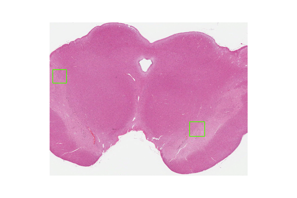
In Parkinson’s disease, dopaminergic neurons in the substantia nigra undergo degeneration, loss, and gliosis formation. Neuronal neuromelanin breaks down and is released from the cells, resulting in depigmentation. Therefore, macroscopically, the substantia nigra loses its normal brown color and turns whitish. Depigmentation also occurs in the locus coeruleus, causing it to macroscopically lose its normal brown color. These macroscopic findings are characteristic of Parkinson’s disease.
In addition to neuronal loss, brain stem-type Lewy bodies are formed in cell bodies. Lewy body formation occurs in the substantia nigra and extensively throughout the brain stem nuclei, including the oculomotor nucleus, locus coeruleus, and dorsal nucleus of the vagus nerve. In the cerebrum, Lewy bodies are also prone to forming within neurons in the Meynert basal ganglia, hypothalamus, and amygdala. Cortical Lewy bodies are also observed in the cerebral cortex, albeit in small numbers. In the peripheral nervous system, Lewy bodies appear in the sympathetic and parasympathetic ganglia, intestinal nerve plexuses, and adrenal glands.
Lewy bodies form not only within cell bodies, but also within neurites. Extracellular Lewy bodies that have been released from cells are also present.
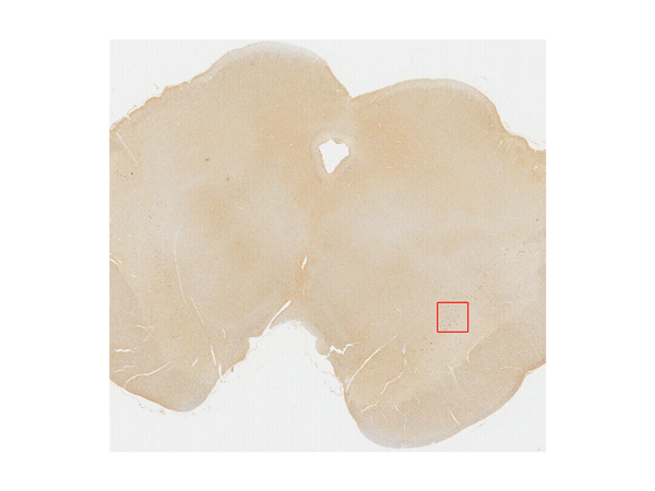
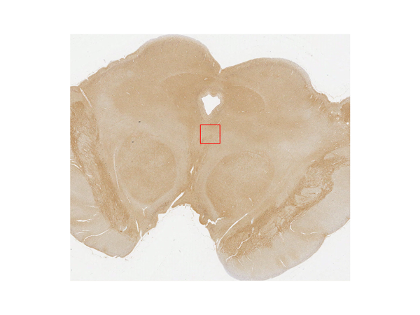
Lewy bodies are positive for α-synuclein and ubiquitin. With α-synuclein and ubiquitin staining, Lewy bodies and some neurites stain positive. These neurites are known as Lewy neurites, or Lewy-related neurites.