

Thank you for using our website.
To provide more stable service, we have migrated our website server starting in June 2025. As a result, the login page URL has changed. Please note the new URL below:
New Login Page URL: https://pathologycenter.jp/kiasma/en-eban-login
If you have bookmarked the previous URL, please update your bookmark to the new URL.
Your existing login account will remain valid, and you can continue to access the site as before.
If you experience any issues—such as being unable to log in or access the site—please contact us at the address below.
【Contact Information】 neuropathology
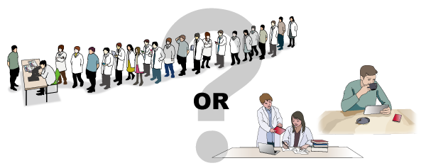
Which observation method is more convenient for you?
You don't even need to say it.
I am pleased to introduce the website about neurohistology and neuropathology created by our laboratory. There are login-required contents and access-free contents. The former is rich in educational contents using brain cutting videos and virtual slides. You can enter the sample room and experience them. If you would like to view all the login-required contents, please apply from the registration form. You can see them for free.
Please view them properly under the suitable combination of device and browser.
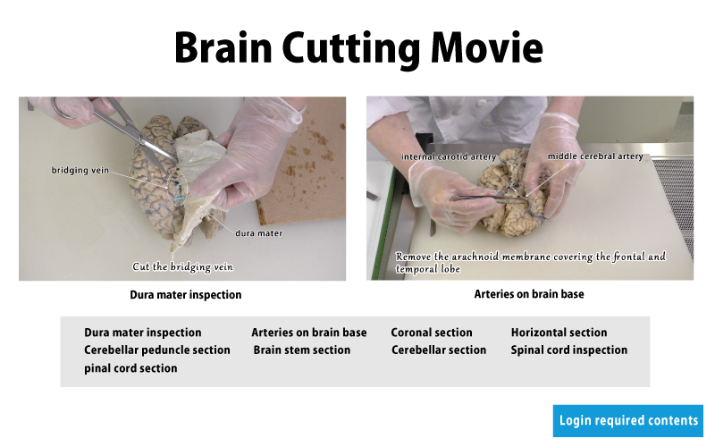
You can watch a lot of videos of brain cutting. The explanation is also written on the video screen, so it is easy to understand.
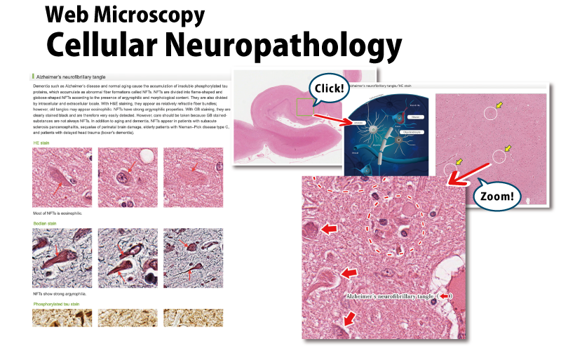
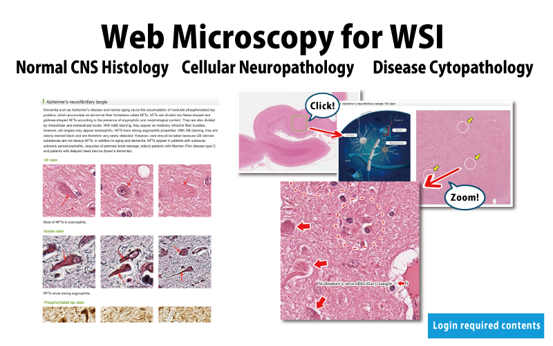
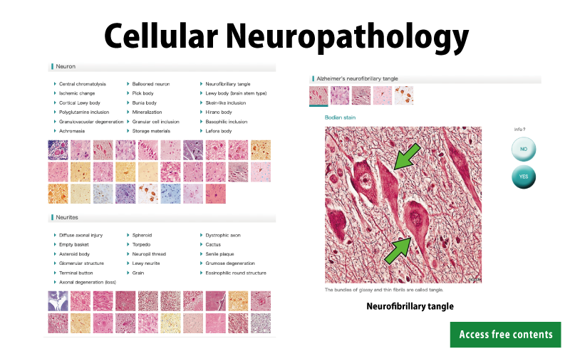
There is a lot of WSIs of pathological structures of neurons and glial cells. The observation area is shown and annotations are also written on the WSI.
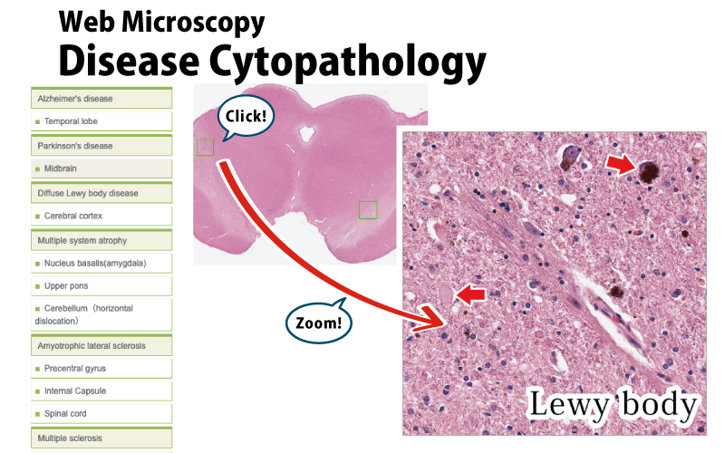
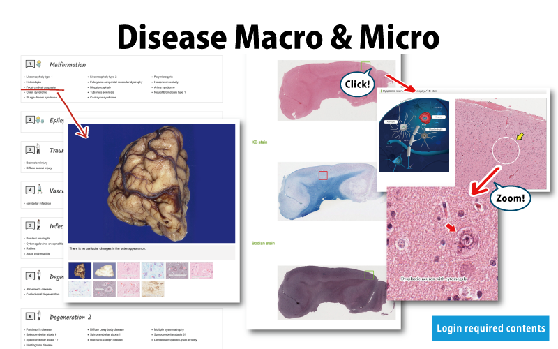
There is a lot of WSIs that can be helpful in diagnosis of neurological diseases. In the future, the number of diseases will increase.
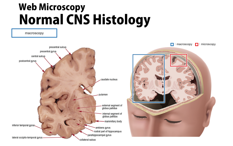
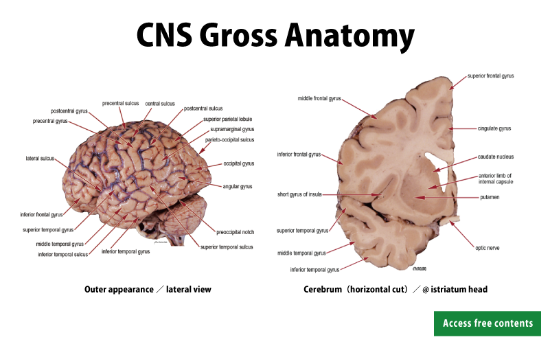
You can view the gross images, the maps using stained specimens, and WSI of the CNS for each location. No better teaching materials of neuroanatomy.
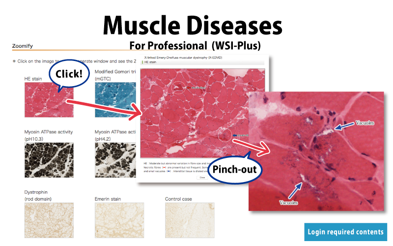
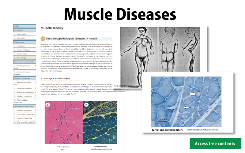
Virtual slides of muscles are very valuable for studying pathology of diseases that primarily and secondarily affect skeletal muscles.

Many of the macro photos posted here were taken at angles that are not found in brain atlas books being published so far. I'm sure you will be interested in them.
Characteristic changes of Macro and Micro are presented by disease category.
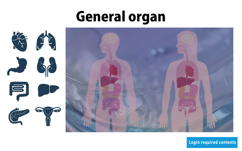
In addition to neuroanatomy, you can also study normal tissues of general organs, including lung, liver, kidney and so on.
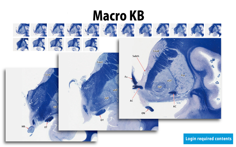
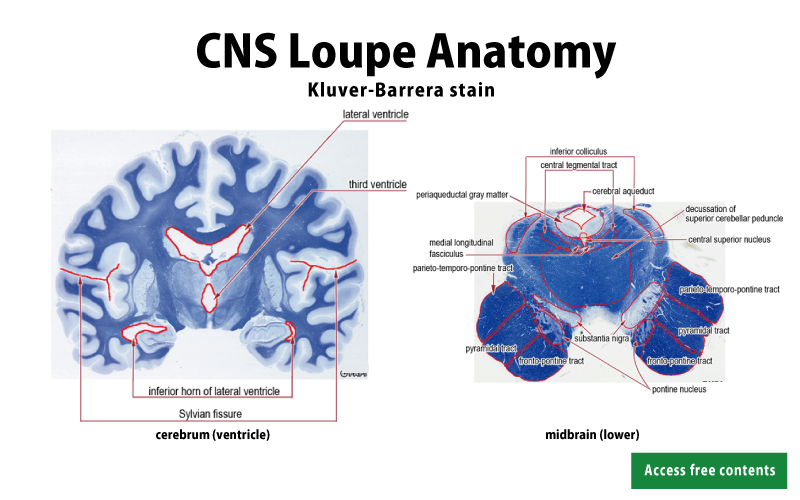
You can study neuroanatomy of various regions with continuous images of KB staining.

Let's study neuroanatomy by looking at the appearance and cut-faces of the CNS. This knowledge is fundamental to neuropathology.
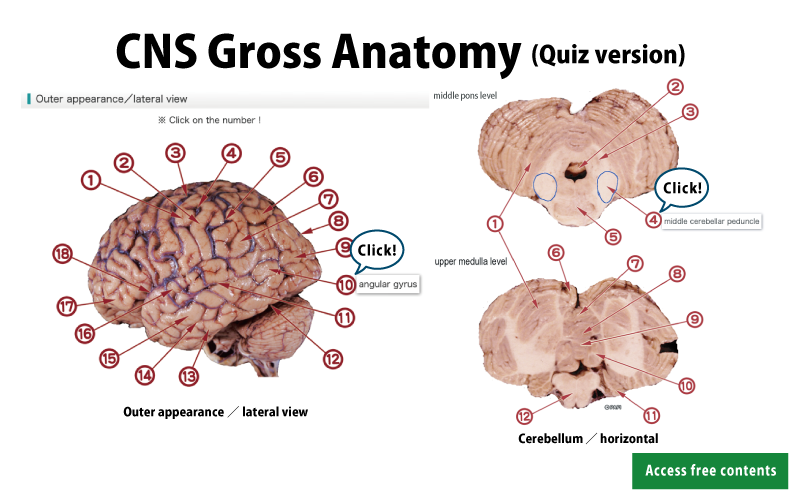
Can you tell the name of each part of the CNS? Challenge the quiz and check your knowledge! Are you okay?
Look at the KB-stained specimens and check each part of the CNS. Let's learn more detailed appearances than macroscopic findings.

We are making a lot of animations as to macroscopic features of neuroanatomy, development and pathology.
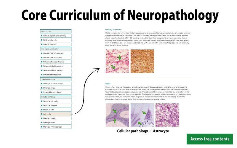
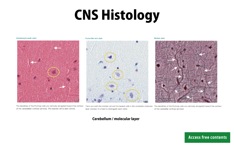
You can check the histology of each part of the CNS. The shape and size of the nerve cell varies depending on the part of the CNS.
You can see a index list of pathological images by cell. It is easy to search the pathological image you want to examine.

The animations explain the pathogenesis of various pathological changes observed with a microscope.
This content explains muscle diseases in an easy-to-understand manner. There are many figures and photos.
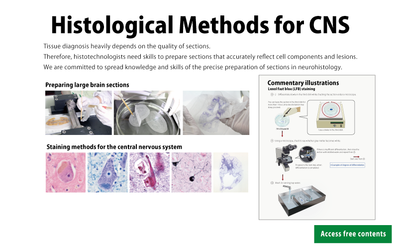
The know-how for making good specimens is explained easily to understand. There are also many photos, illustrations, and videos.